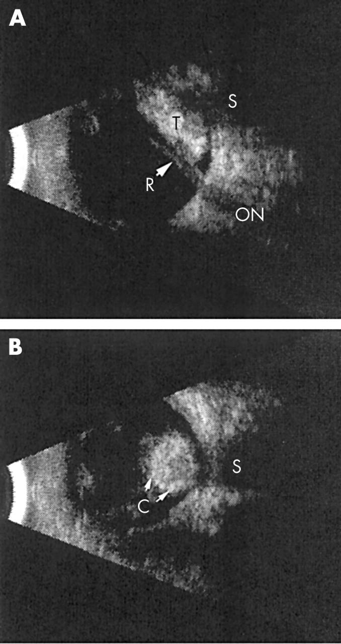
Figure 1.

B-scan ultrasonography. (A) A longitudinal view of the right eye demonstrates a tumour posterior to a closed funnel retinal detachment. (B) A transverse view of the eye demonstrates a rounded, densely calcified tumour. R, retina; S, shadow in orbit; C, calcification; T, tumour; ON, optic nerve.
