
Figure 2.
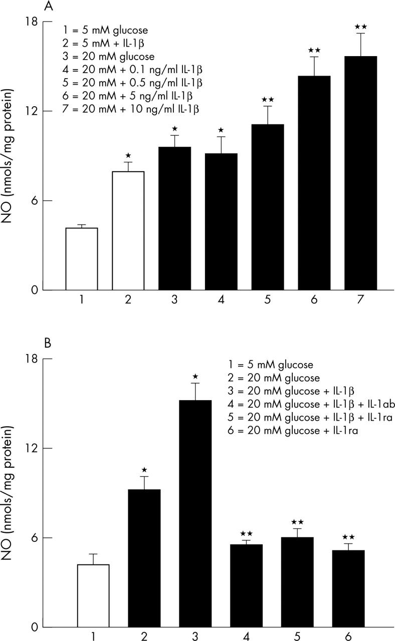
Effect of IL-1β on glucose induced NO in retinal endothelial cells. (A) NO content was measured in the endothelial cells incubated in 5 mM or 20 mM glucose for 3 days in the presence or absence of recombinant IL-1β. The data shown are obtained from the cells incubated in 5 mM glucose in the presence of 10 ng/ml IL-1β, and incubated in 20 mM glucose containing four different concentrations of IL-1β. (B) To confirm the effect of IL-1β on glucose induced NO, the cells were also incubated with 50 ng/ml IL-1ab or 100 ng/ml IL-1βra for three days. Each measurement was performed in duplicate. *p<0.05 compared with 5 mM glucose, and **p<0.05 compared with 20 mM glucose.