
Figure 4.
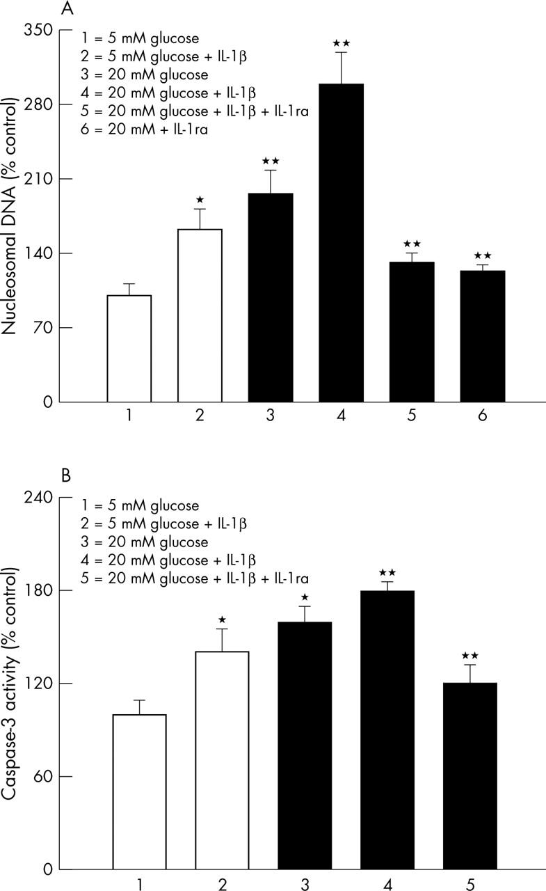
Effect of IL-1β on capillary cell death. (A) Apoptosis was measured by performing ELISA for cytoplasmic histone associated DNA fragments using an assay kit from Roche Diagnostics. The values obtained from the cells incubated in 5 mM glucose are considered 100%. The concentration of IL-1β in the incubation medium was 10 ng/ml, and the incubation time was five days. The values obtained were adjusted to the total DNA. (B) Effect of IL-1β on the activation of apoptosis execution enzyme caspase-3 was determined in the cells incubated in 5 mM and 20 mM glucose in the presence or absence of 10 ng/ml IL-1β for 5 days using the fluoregenic substrate DEVD-AFC. The fluorescence signal emitted was quantified at excitation and emission wavelengths of 400 nm and 505 nm respectively. Each experiment was repeated with four different cell preparations, and measurements made in duplicate. The values obtained with 5 mM glucose were considered as control values. *p<0.05 and **p<0.05 compared with 5 mM and 20 mM glucose, respectively.