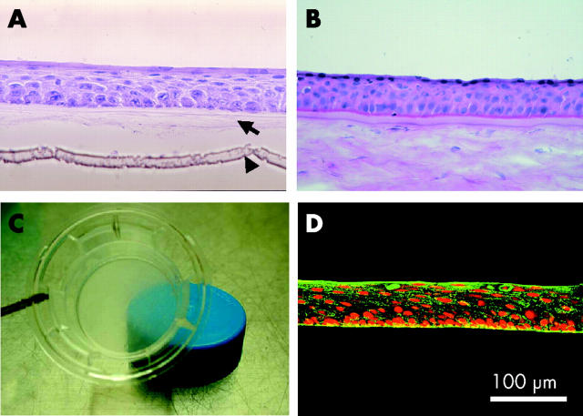
Figure 1.
Cultivation of oral epithelium on amniotic membrane. Light micrographs of cross sections of (A) epithelial cells from oral biopsy samples of patient 2 cultivated on amniotic membrane and (B) normal corneal epithelial cells stained with haematoxylin and eosin. The arrow in (A) points to the denuded amniotic membrane; the arrowhead identifies the membrane of the culture insert (original magnification ×400). (C) The culture insert retained its transparency as indicated by the translucence of the blue cap beneath the culture insert. (D) The cultured oral epithelial cells are immunohistochemically stained for keratin 3 (green). Their nuclei are positive for propidium iodide (red).

