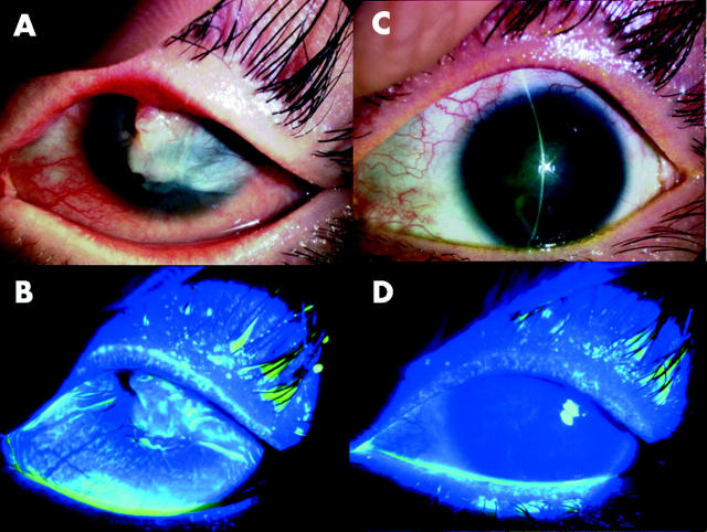
Figure 3.
Transplantation to a patient with SJS of autologous oral epithelial cells cultivated on amniotic membrane (patient 4). Representative slit lamp photographs taken before transplantation without (A) and with fluorescein (B). The photographs in (C–D) were taken at the last follow up visit without (C) and with fluorescein (D). Before transplantation, the eye manifested inflammatory subconjunctival fibrosis with neovascularisation, conjuntivalisation, and severe symblepharon. At the last follow up visit, the corneal surface was stable without defects.

