
Figure 1.
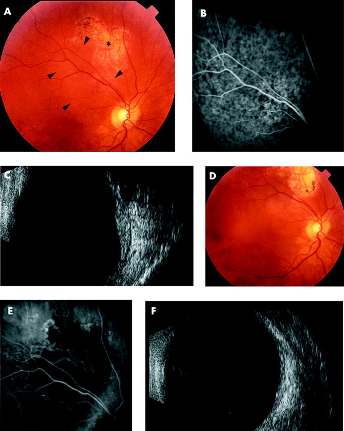
(A) Fundus photograph of the right eye showing a circumscribed choroidal haemangioma (arrowheads). The tumour was unsuccessfully treated with transpupillary thermotherapy (*). (B) Indocyanine green angiogram showing choroidal hypervascularity and hyperfluorescence due to choroidal haemangioma. (C) B-scan ultrasonograph of a dome shaped choroidal lesion and high internal reflectivity (thickness 2.5 mm). (D) Post-treatment fundus appearance showing regression of haemangioma. Areas of retinal pigment epithelial stippling and atrophy are present in the treated areas. (E) Post-treatment indocyanine green angiogram demonstrating that the choroidal haemangioma is replaced by an area of hypovascular choroid. (F) Post-treatment B-scan ultrasonograph showing marked regression of the choroidal haemangioma.