
Figure 4.
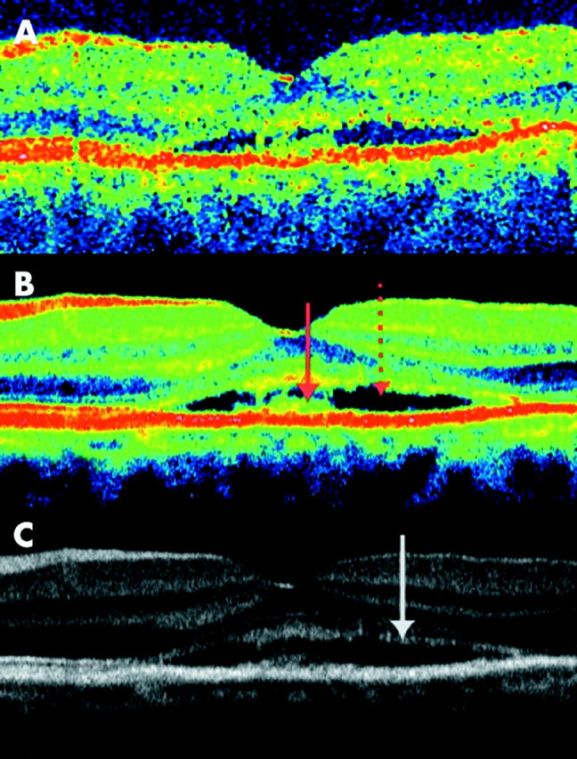
Transfoveal optical coherence tomograms (OCT3, horizontal orientation, nominal width 5 mm) of the left eye of a 50 year old woman with 6 months complaint of blurred vision and photograpic (not shown) and clinically evidence of central serous retinopathy. (A) Conventional single scan image. (B and C) Averaging of eight images. The serous detachment and debris at the inner surface of the RPE are shown with arrows on the colour image. A serous detachment is seen at the level of the middle hyperreflective band, bordered with debris both on the posterior border of the neuroretina and the anterior border of the RPE. As seen on the greyscale image, both the hyperreflective lines from the outer limiting membrane and the junctions of inner and outer photoreceptor segments (arrow) are of lower intensity but otherwise comparable with the healthy eye.