
Figure 2.
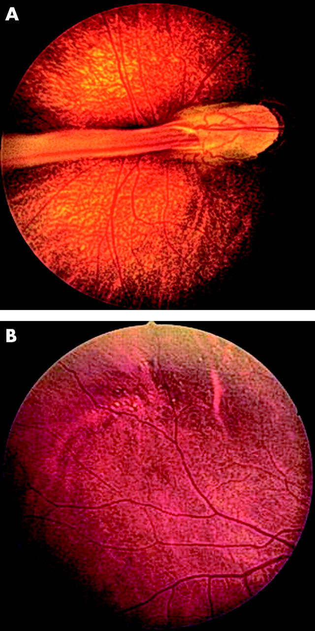
Clinical pictures of family members showing classic features of FEVR. (A) Fundus photograph of the proband’s (V:6) optic nerve and macula in the right eye taken at 7 months. Virtually all of the retinal vessels are drawn up into a “comet”-shaped retinal fold which extends from the optic nerve to a large fibrovascular mass in the temporal retinal periphery. (B) Fundus photograph of the proband’s father’s (IV:6) temporal peripheral retina. The optic nerve and macula in this individual appeared normal but examination of the peripheral retina revealed the presence of multiple exudates (yellow dots).