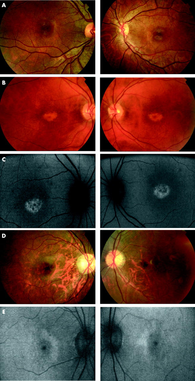
Figure 2.

(A) Case 7. Fundus photographs showing bilateral macular atrophy, left worse than right. (B) Case 5. Fundus photographs showing bilateral “bull’s eye maculopathy (BEM)-like” RPE changes. (C) Case 5. Fundus autofluorescence imaging showing bilateral central macular areas of markedly increased AF surrounded by a ring of relative decreased AF. (D) Case 1. Fundus photographs showing bilateral mild macular atrophy with mild temporal optic nerve pallor. (E) Case 1. Fundus autofluorescence imaging showing bilateral perifoveal rings of relative increased AF.
