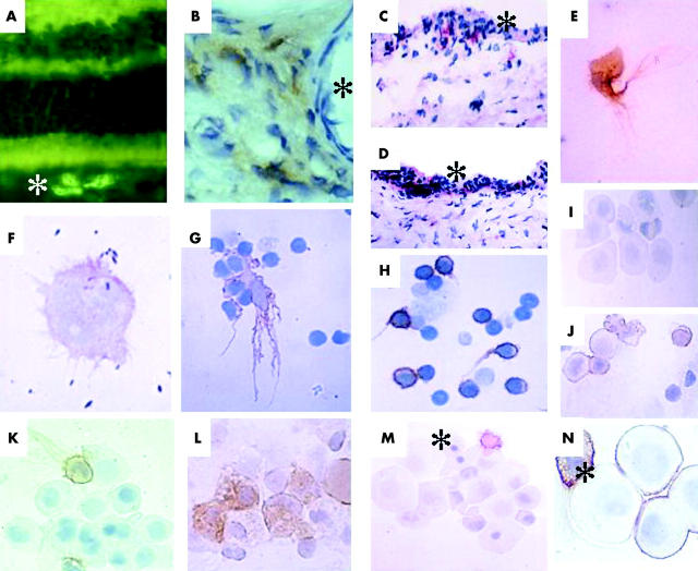
Figure 1.
Immunohistochemistry of choroidal dendritic cells in sections from normal rat eyes and in cytospin preparations after culture in vitro (see Methods). (A) Immunofluorescence of section of rat eye stained for MHC class II (OX6); note MHC class II positive cell in the choroid (*). Photoreceptors and bipolar cells also show autofluorescence caused by retinal chromophores (see Brissette-Storkus et al9). (B) Immunoperoxidase stained section of choroid showing brown stained MHC class II (OX6) positive dendritic cells surrounding a vein (*marks the lumen). (C) APAAP stain of choroid-retinal pigment epithelium (RPE) explant with ED2+ (red) cells present in the RPE layer (*). (D) APAAP stain of choroid-RPE explant showing OX62+ (red) cells present in the RPE layer (*) and in the choroidal stroma. (E) Cytospin preparation of immunoperoxidase-stained culture of rat MHC class II+ dendritic cell showing extensive veiled phenotype. (F) Cytospin APAAP preparation of cultured human dendritic cell showing moderate positivity for B7 antigen; the rod-like structures also present in the figure are photoreceptor fragments. (G) Cytospin preparation of large veiled MHC class II positive (immunoperoxidase) rat dendritic cell clustered with T cells in co-culture (see Methods). (H) Cytospin preparation of small intensely MHC class II positive dendritic cells with long tails forming small T cell clusters after co-culture. (I) Cytospin preparation of large round low densitiy MHC class II negative choroidal cells forming clusters in culture. (J) similar preparation to (I) stained for ED7 antigen (immunoperoxidase). (K) Cytospin preparation of large cell cluster harvested from choroid-RPE explant by washing, and showing two MHC class II positive dendritic cells in contact with the MHC class II negative large cells. (L) Cytospin preparation of a cluster of large cells freshly harvested from collagenase digested choroidal-RPE explant and showing strong ED2 positivity in a proportion of the cells. (M) Cytospin preparation of a cluster of large cells some of which contain pyknotic nuclei (*), in contact with a single MHC class II positive dendritic cell. (N) Similar preparation to (M) but at higher magnification showing a single ED7 positive dendritic cell (*) in contact with four large cells, three of which are also ED7 positive.

