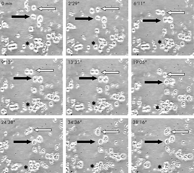
Figure 3.
Time lapse video microscopy. Single shots from a video time lapse photographic sequence of choroidal dendritic cells in culture. Black arrow indicates a motile but non-translocating cell while white arrow identifies two translocating cells which contact and separate from the non-translocating cell. (*) identifies a large motile cluster of cells.

