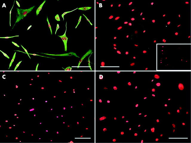
Figure 3.
Immunostaining of cultured cells derived from pterygium fibrovascular tissue. All cells are uniformly positive to vimentin (A), while some are positive to α-SMA (B), negative control to α-SMA in conjunctivochalasis fibroblast (inset) but are negative to desmin (C) and caldesmon (D). The bar represents 50 μM.

