
Figure 4.
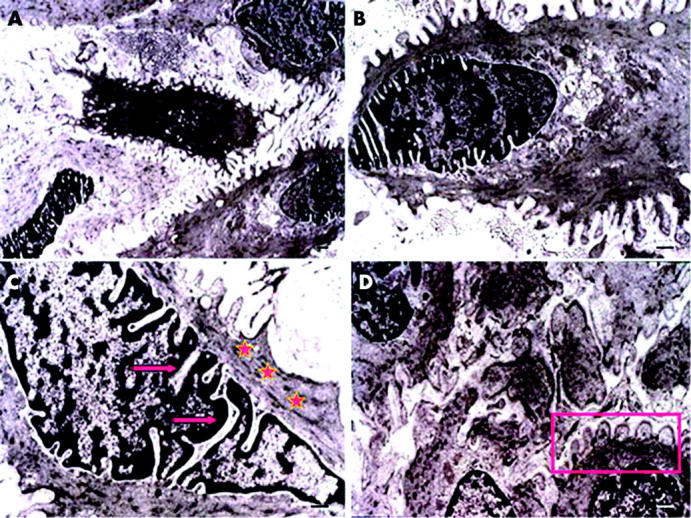
Ultrastructural features of cells in pterygium fibrovascular tissue. Cells show intracytoplasmic bundles of microfilaments (stars) arranged in parallel to the axis of the cell at the periphery (A), numerous nuclear indentations (arrows) and deep folds that give indirect evidence of contraction (C), and active pseudopodal activity (B and D).