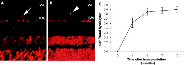
Figure 4.
The kinetics of the hyalocytes. Chimeric mice were used to analyse the hyalocytes’ origin and regeneration at 0, 4, 6, 7, and 12 months after BM transplantation by fluorescent microscopy. The PI+ nuclei associated GFP− cells, indicating recipient derived hyalocyte, were seen in the mouse right after BM transplantation (A). The donor derived (GFP+) hyalocytes were observed 6 months after BM transplantation (B). The time dependent ratio of GFP+ hyalocytes in the chimeric mice (C) (original magnification ×400).

