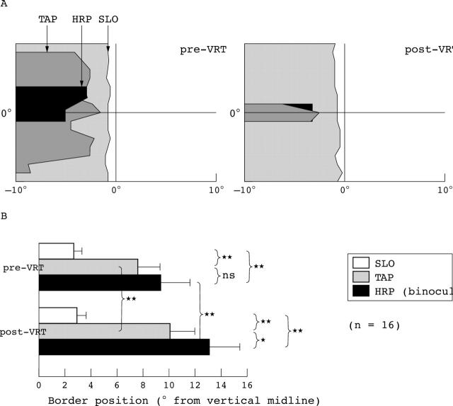
Figure 1.
This graph (adapted from Sabel et al4) displays the visual field border position in the right eye as assessed by the three perimetric tests. (A) The results of patient CH where grey areas represent the area of the defect. A mismatch in perimetric fields was noted even before therapy. After VRT, the HRP and TAP border shifted away from the vertical meridian whereas the SLO border remained roughly in the same position, exaggerating the border mismatch. (B) Shows the absolute visual field border for SLO, TAP, and HRP in the central 10° region in degrees of visual angle from the 0 vertical meridian before and after VRT (mean (SEM)). Whereas the SLO border was almost identical pre-VRT compared with post-VRT, the HRP and TAP borders were not only significantly different before VRT (mismatch), but also both shifted significantly after VRT, producing a visual field enlargement.4

