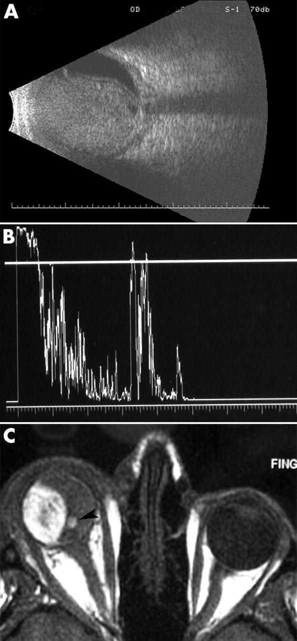
Figure 1.

(A) B-scan ultrasound demonstrated a large intraocular tumour with scleral thickening and retrobulbar oedema. There was almost no intrinsic vascularity noted within the tumour. No extrascleral tumour extension was noted. (B) A-scan ultrasonography of the right eye showed relative low internal reflectivity. (C) MRI of the orbits was significant for an intraocular tumour that displayed high signal intensity on T1 weighted images. On T2 weighted images it displayed low signal intensity. Note the small eccentric collar-button (arrow). There was no evidence of extraocular tumour extension.
