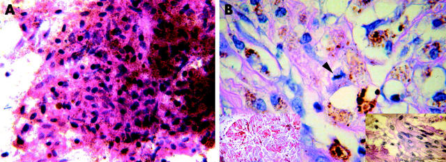
Figure 2.
(A) The cytological preparations from FNAB showed very small, pigmented dendritic and epithelioid cells with small dark oblong nuclei. No mitoses or necrosis was present. Some of the very deeply pigmented cells seen were melanophages. These cells were naevus-like, there were no cells that were large enough or that had appropriate nuclear morphology to be called melanoma (original magnification ×100). (B) Histopathology of the enucleated eye showed a necrotic tumour (left inset; original magnification ×100). Histopathology demonstrated spindle and small epithelioid melanoma cells (right inset; original magnification ×100) and a melanoma with a mitotic figure in the centre (arrow). These findings suggested malignant change (original magnification ×100).

