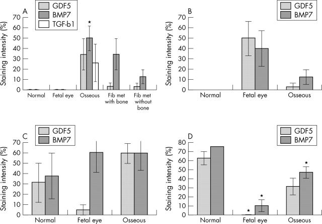
Figure 4.
Staining intensity of GDF-5 and BMP-7 in (A) RPE; (B) retina; (C) ciliary epithelium; and (D) cornea of normal adult and fetal eyes, and eyes with osseous metaplasia. The staining intensity of TGF β1 is only evaluated in the RPE (A), and includes eyes with RPE fibrous metaplasia without bone formation.

