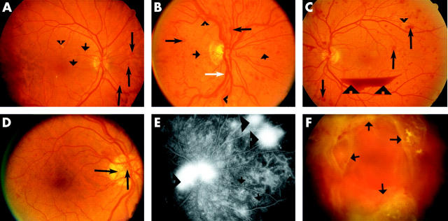
Figure 2.
Colour fundus photography (CFP) and fluorescein fundus angiography (FFA) showing different features of diabetic retinopathy (DR). (A) An eye with mild non-proliferative diabetic retinopathy (NPDR) presented with microaneurysms (short arrows), haemorrhages (long arrows), as well as hard and soft exudates (arrowhead). (B) An eye with severe NPDR showing a greater number of microaneurysms (short arrows), haemorrhages (long arrows), and also venous abnormalities such as venous dilatation (white arrow) and tortuosity (arrowheads). (C) and (D) Eyes with high risk proliferative diabetic retinopathy (PDR). Haemorrhages (long arrows), hard exudates (small arrowhead), venous beading on the inferior arcade, new vessels (hallmark for PDR) and preretinal haemorrhage (large arrowheads) are present in (C) and new vessels at the disc (long arrows) are seen in (D). (E) A late phase FFA of an eye with PDR showing leakage from the new vessels (arrowheads) and small spots of hyperfluorescence representing microaneurysms (arrows). Dark patches near the right edge are zones of capillary non-perfusion. (F) Eye with advanced PDR showing extensive fibrovascular proliferation (arrows).

