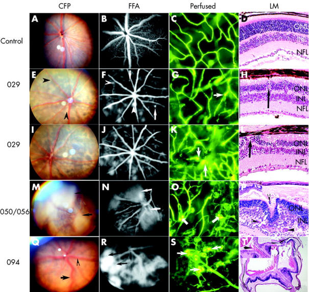
Figure 3.

Characterisation of the different transgenic mouse lines by colour fundus photography (CFP) (A, E, I, M, and Q), fluorescein fundus angiography (FFA) (B, F, J, N, and R), fluorescence micrographs of flat mounted, fluorescein labelled dextran perfused eyes (C, G, K, O, and S), and light micrographs (LM) of haematoxylin and eosin stained paraffin sections (D, H, L, P, and T). C57Bl/6 control mouse eye at 4 weeks (A–C) and at 3 weeks postnatal (D). Line 029 transgenic mouse eye at 4 weeks (E–G) and at 3 weeks (H) postnatal. Line 029 transgenic mouse eyes at 12 weeks (I–J) and at 10 weeks (K–L) postnatal. Line 050/056 transgenic mouse eye at 4 weeks (M–O) and at 3 weeks (P) postnatal. Line 094 transgenic mouse eyes at 4 weeks (Q–S) and at 3 weeks (T) postnatal. Arrowhead in (F) demonstrates focal hyperfluorescence which corresponds to pale lesions in (E). Arrows in (N) and (R) point to intense and extensive fluorescein leakage. The arrows in (G), (K), (O), and (S) point to focally dilated vessels (G), microaneurysm (K), and glomerular tufts of hyperfluorescence (O and S). The arrows in (H), (L), (P), and (T) point to new vessels. Arrowhead and arrow in (Q) show venous dilatation and tortuosity, respectively.
