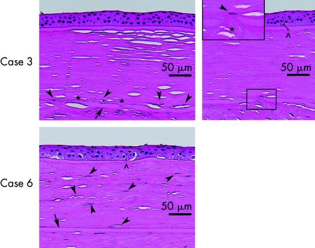
Figure 2.
The central portion of the lenticules and host corneas from cases 3 and 6. Arrow indicates Bowman’s layer of the host cornea. Arrowheads indicate keratocytes repopulated in the lenticules. Asterisk denotes a break observed in Bowman’s layer of host. The symbol ∧ indicates disruption of Bowman’s layer in the lenticule. In case 3, a spindle cell appeared to extend through the break in Bowman’s layer from the host stroma to the lenticule. The repopulated keratocytes were situated adjacent and right above the host Bowman’s layer. In case 6, keratocyte repopulation was observed throughout the anterior and posterior regions of the lenticules (bar indicates magnification).

