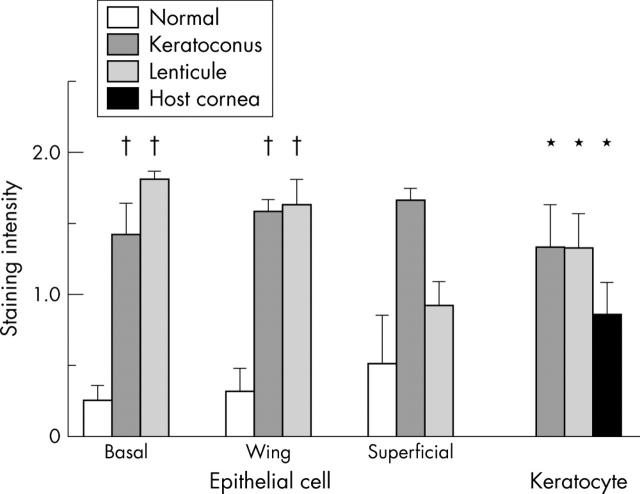
Figure 4.
Staining intensity for Sp1 in the corneal epithelial cells and keratocytes in the lenticules (n = 12), host stromas (n = 12), and normal human (n = 4) and keratoconus (n = 3) corneas as scored by three masked observers. The scores were analysed by Mann-Whitney U tests. *p<0.01 compared with normal human specimens; †p<0.05.

