Figure 5.
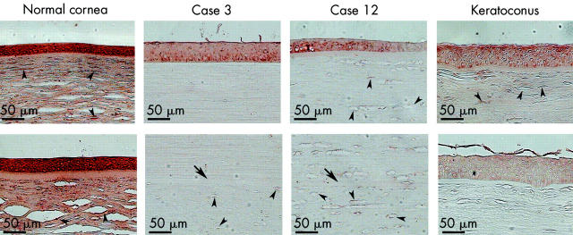
Immunostaining for α1-proteinase inhibitor in corneas from 83 year old (top) and 78 year old (bottom) normal individuals, and a 70 year old keratoconus patient, as well as lenticules of patients after epikeratoplasty for keratoconus (cases 3 and 12). Arrow indicates Bowman’s layer of the host cornea. Arrowheads indicate keratocytes. Note that the intensity of the brown positive staining is lower in the epithelium, keratocytes, and stromal lamellae in the lenticules and host corneas than that in normal corneas. Staining is also weaker in keratoconus corneas compared to normal controls (chromagen 3-3’ diaminobenzidine). Image analysis confirmed significant differences in labelling intensity between normal control (epithelium: basal: 153.5 (3); wing: 141 (3); stromal cell: 179 (3); stromal extracellular matrix (ECM) 72 (5)) and epikeratoplasty case 3 (epithelium: basal: 63 (8) (p<0.0000001); wing: 53 (4) (p<0.000001); stromal cell in lenticule: 31 (6) (p = 0.000001), stromal ECM in lenticule: 14 (2) (p<0.000001); stromal cell in host: 19 (2) (p<0.00001), stromal ECM in host: 12 (1) (p<00001)) and keratoconus (epithelium: basal: 43 (4) (p<0.0000001); wing: 49.3 (10) (p<0.00000001); stromal cell: 37 (8) (p<0.000001); stromal ECM 12.5 (3) (p<0.00001)). Image analysis for case 12 also showed highly significant differences from control.

