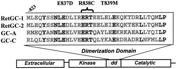
Figure 1.
Location of the dominant cone–rod dystrophy amino acid changes in the dimerization domain (dd) of RetGC-1. A comparison of the sequences of several membrane GCs is also shown, including RetGC-2 (GC-F), the atrial natriuretic peptide receptor (GC-A), and the heat-stable enterotoxin receptor (GC-C). The amino acid residues in boldface are conserved among these membrane GCs.

