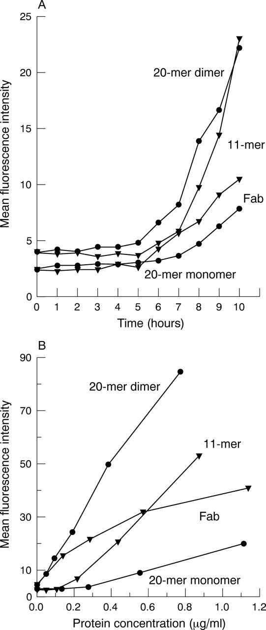
Figure 5.

Penetration of different antibody fragment formats across the perfused pig cornea. (A) Mean fluorescence intensity (as measured by flow cytometry on rat thymocytes) of perfusate sampled from representative corneas treated topically with scFv 20-mer monomer and dimer, scFv 11-mer dimer, and Fab monomer. (B) Titration series of purified protein for each antibody fragment format. Dilutions of purified fragment were tested for reactivity against rat thymocytes by flow cytometry.
