
Figure 3.
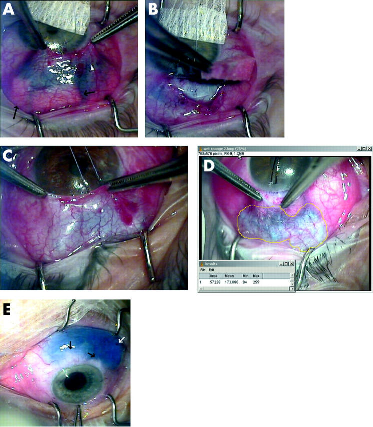
(A) Effect of adding mitomycin C (MMC) with 0.01% trypan blue to preplaced dry sponges during trabeculectomy. Arrows show posterior sponges with no trypan blue staining indicating no uptake of MMC. (B) One of the posterior sponges is removed, confirming no MMC uptake. Trypan blue can be seen staining the anterior operative field up to the limbus. (C) Effect of using sponges presoaked in MMC with 0.01% trypan blue seen through thick (C) and thin (D) tenon’s capsule. The treatment area now corresponds better to the area of sponge placement. (D) The area of treatment has been measured (area in pixels using Image J, (NIH, Bethesda, MD, USA). Note no inadvertent staining with trypan blue around the limbus. (E) Subconjunctival injection of 45 mg/ml 5-FU with 0.01% trypan blue after cataract surgery on an eye with a 680 day old trabeculectomy bleb. Black arrows show subconjunctival fibrous bands. White arrow shows point of conjunctival entry with ooze of blood but no trypan blue.