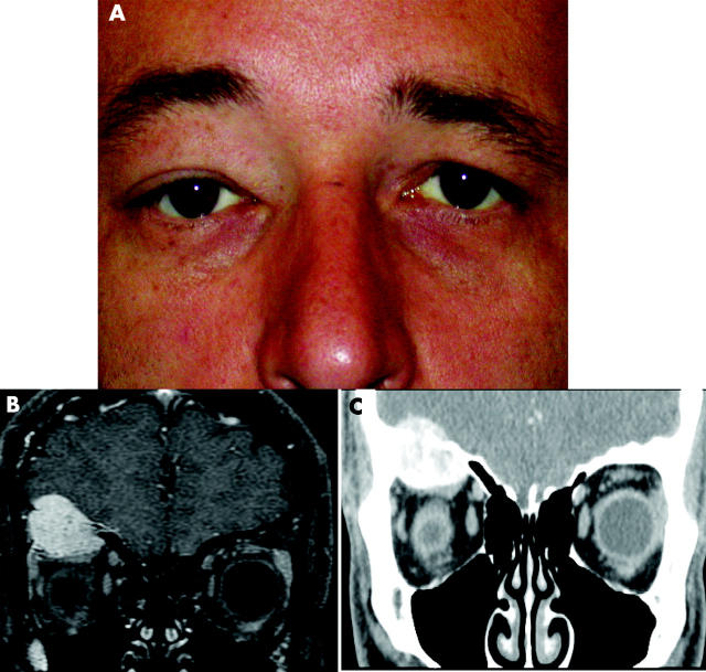
Figure 1.
(A) A 39 year old patient showing proptosis and ptosis in the right eye. (B) Gadolinium enhanced coronal T1 fat saturated image through the orbits demonstrates an intraosseous mass in the right orbital roof, with intraorbital and intracranial extension. The intracranial portion was completely extra-axial, with associated dural involvement, as indicated by the thickened and enhancing dura adjacent to the dominant intracranial component. (C) Contrast enhanced coronal computed tomography (CT) image through the orbits demonstrates an intraosseous mass in the right orbital roof, with intraorbital and intracranial extension. Its heterogeneous appearance is the result, in part, of scattered calcifications throughout the mass. Mass effect upon the superior extraocular muscle group is evident.

