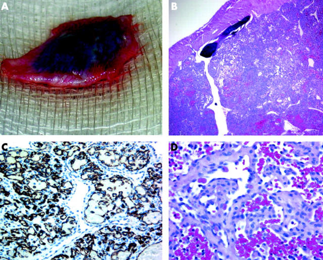
Figure 2.

(A) Gross tumour mass showing involved resected dura. (B) HPE: 8×4 magnification showing thin walled blood vessels and osteoblastic activity of intraosseous cellular capillary haemangioma. (C) 6×40 magnification with CD 34 positivity confirming vascular origin. (D) 6×40 dural involvement by capillary haemangioma.
