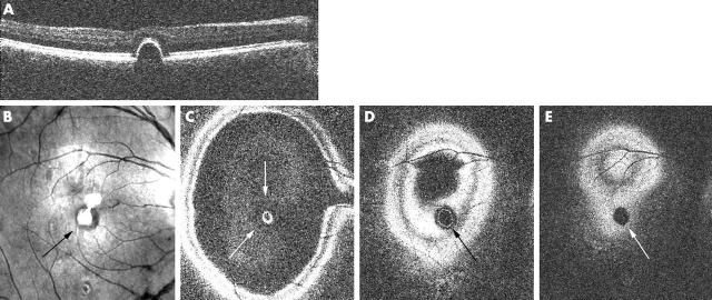
Figure 3.
Retinal pigment epithelium (RPE) detachment (observed in an inactive CSR patient) as seen on OCT C-scan. (A) En face OCT B-scan showing the RPE detachment. (B) Confocal C-scan image showing a change in the reflectivity of the retina in the area corresponding to the RPE detachment (arrow; central bright white spots are artefacts from the focusing lenses within the imaging device system). (C–E) Consecutive OCT C-scans showing the circular area of the PED, with outer retinal elevation (white arrows in C), the inner circle (black arrow in D) corresponding to the RPE detachment, and the dark circular area (white arrow in E) corresponding to the area underneath the PED, caused by its shadowing effect.

