Figure 4.
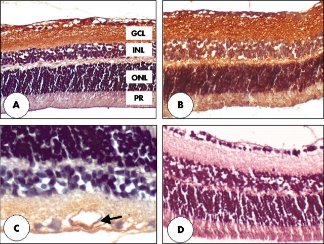
Immunohistochemistry of gremlin in retina of diabetic and non-diabetic mice. The nerve fibre layer (NFL), ganglion cell layer (GCL), and inner plexiform layers (IPL) of both non-diabetic (A) and diabetic (B) mice show strong immunoreactivity. Diabetic animals demonstrate gremlin immunoreactivity in the outer retina (B) and also at the level of the vascular endothelium (arrow), especially noticeable in the large retinal vessels (C). Primary antibody omission controls show no apparent deposition of DAB reaction product (D). Original magnifications: ×200 (A, B, and D). ×400 (C). The outer nuclear layer (ONL) and photoreceptors (PR) are also labelled.

