
Figure 2.
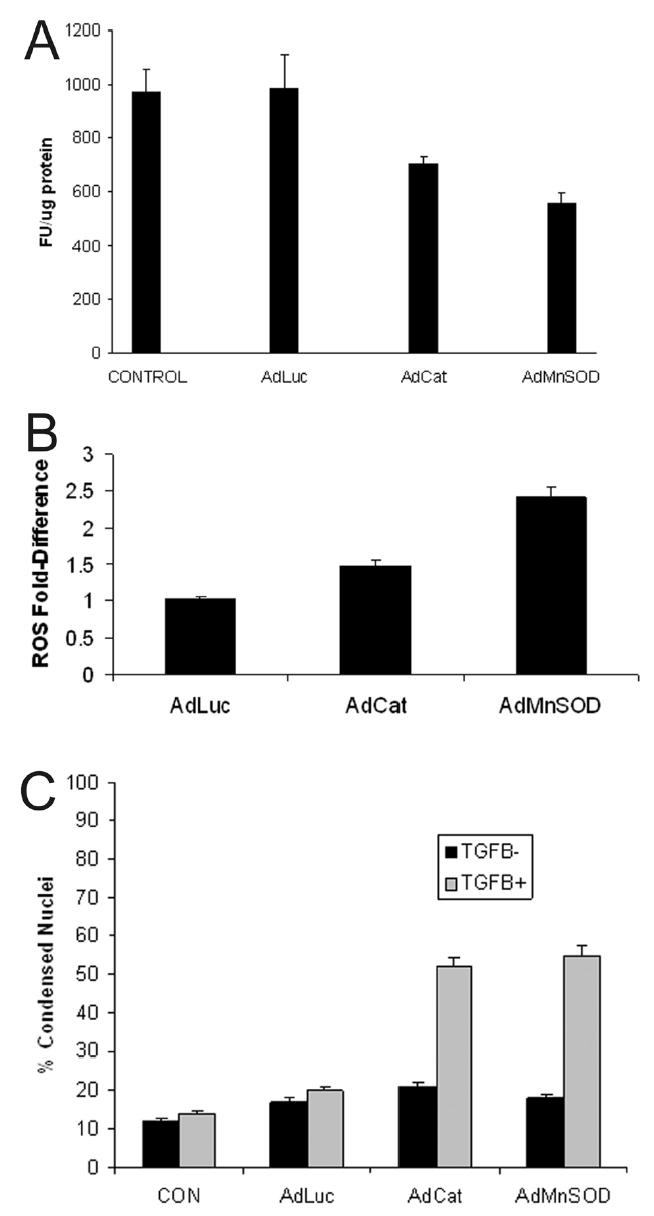
(A) Cirrhotic hepatocytes were transfected with adenoviruses (MOI 100) expressing luciferase (AdLuc; control), catalase (AdCat), or MnSOD (AdMnSOD) for 24 hours and ROS activity was fluorometrically determined using DCF. Some hepatocytes were untreated with adenovirus (control). Transfection with AdCat and AdMnSOD decreased significantly ROS formation (p<0.05 vs. control). (B) Cirrhotic hepatocytes were transfected with adenoviruses (MOI 100) expressing luciferase (AdLuc), catalase (AdCat), or MnSOD (AdMnSOD) 24 hours prior to TGFβ (5 ng/ml) treatment and incubated with DCF. Generation of ROS, expressed as fold-difference, was fluorometrically determined at 90 minutes following TGFβ administration. The adenoviruses expressing the antioxidants, catalase and MnSOD permitted a ROS burst at 90 minutes, indicating ROS responsiveness to TGFβ administration in cirrhotic hepatocytes. (C) The percent of condensed nuclei, indicative of morphologic apoptosis, was determined at 48 hours in cirrhotic hepatocytes in response to treatment with or without TGFβ (5 ng/ml) after pretreatment for 24 hours with adenoviruses (100 MOI) expressing luciferase (AdLuc), catalase (AdCat), or MnSOD (AdMnSOD). Transfection of cirrhotic hepatocytes with adenoviruses expressing the antioxidants, catalase and MnSOD, followed by treatment with TGFβ increased significantly the percent of apoptotic hepatocytes compared to control (p<0.05 vs. control).