
Figure 5.
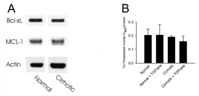
(A) Whole tissue lysates were prepared from normal and cirrhotic livers and levels of Bcl-xL and MCL-1 were determined by Western blot analysis. Actin levels were also immunoblotted to ensure an equal protein loading. (B) Hepatocytes isolated from normal and cirrhotic livers were incubated in HDM in the presence or absence of 5 ng/ml TGFβ, as described in MATERIALS and METHODS. After 48 hours, cell lysates were prepared and total protein tyrosine phosphatase activity was determined spectrometrically.