Figure 6.

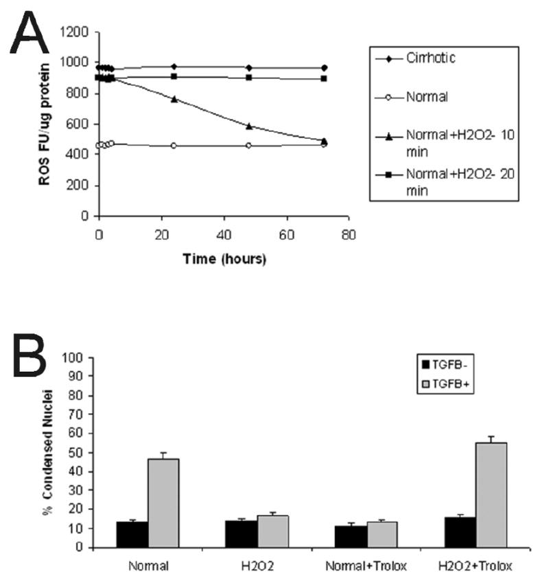
(A) ROS fluorescent units (FU) were determined over time in normal, cirrhotic, and normal-H2O2-converted hepatocytes. Normal hepatocytes (open circles) have a low basal level of ROS activity whereas cirrhotic hepatocytes (black diamonds) have a high steady-state level of ROS activity. Normal hepatocytes exposed to 10 μM H2O2 for 10 minutes (black triangles) failed to maintain a sustained ROS response. However, normal hepatocytes exposed to 10 μM H2O2 for 20 minutes (black squares) demonstrate sustained increased ROS activity similar to basal cirrhotic hepatocytes over the study period. These hepatocytes had 97% viability (data not shown). (B) The percent of condensed nuclei was determined in normal and H2O2-converted hepatocytes at 48 hours after pretreatment (or not) with trolox (2 μM) followed by treatment with or without TGFβ (5 ng/ml). Treatment with H2O2 alone did not increase apoptosis, and, similar to cirrhotic hepatocytes, H2O2-converted cells were resistant to TGFβ -induced apoptosis at 48 hours. However, H2O2-converted hepatocytes that were pretreated with trolox prior to TGFβ exposure underwent apoptosis similar to normal hepatocyte controls and antioxidant-treated cirrhotic hepatocytes.
