
Figure 2.
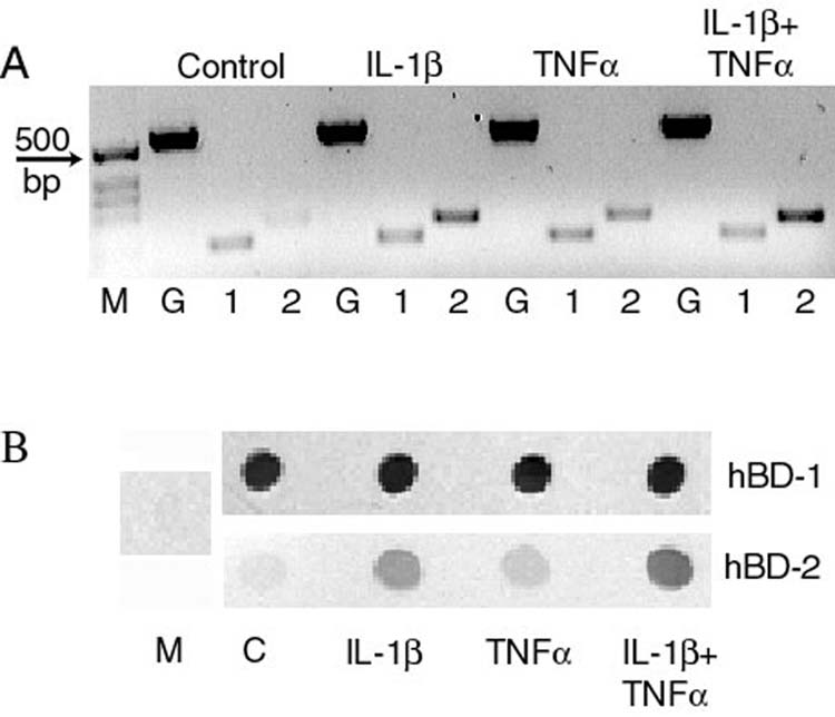
Effect of cytokines on β-defensin expression by primary cultured corneal epithelial cells. Cells were treated with 10ng/ml IL-1β, TNFα or both for 24 hours then RT-PCR (panel A) was used to study defensin mRNA expression and immunoblotting (panel B) was used to study peptide secretion in to the culture media. Panel A: M = size markers, G = housekeeping gene glyceraldehyde-3-phospahte dehydrogenase, 1 = hBD-1, 2 = hBD-2. Panel B: M = culture media that had not been in contact with cells, C = supernatant from control cells. Expression of hBD-1 is constitutive whereas hBD-2 is upregulated by IL-1β and TNFα. (Reprinted from McDermott AM, Redfern RL, Zhang B, et al. Defensin expression by the cornea: multiple signaling pathways mediate IL-1β stimulation of hBD-2 expression by human corneal epithelial cells. Invest Ophthalmol Vis Sci, 2003;44:1859-65, with permission of Association for Research in Vision and Ophthalmology.)