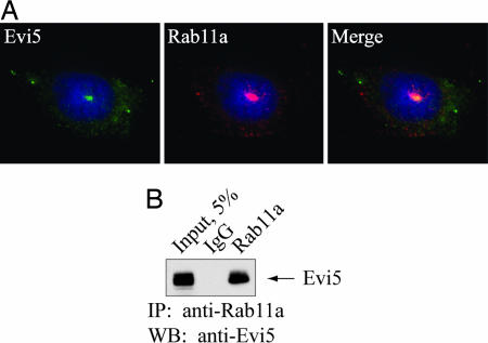
Fig. 2.
Evi5 and Rab11 bind in vivo. (A) Endogenous Evi5 and Rab11a proteins colocalize in the pericentriolar region of the cell. U2OS cells were processed for immunofluorescence analysis by using affinity-purified anti-Evi5 and anti-Rab11 antibodies, as well as Hoechst to mark DNA. (B) Endogenous Evi5 and Rab11a proteins form a complex in vivo. Equal amounts of 293T cell lysate were subjected to immunoprecipitation by using either rabbit IgG or antibody against Rab11a. Captured immune complexes were eluted with sample buffer and subjected to SDS/PAGE and immunoblotting with anti-Evi5 antibody.

