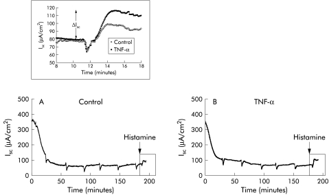
Figure 8.
Changes in short circuit current (Isc) across mouse distal colon over a three hour period of measurements for two typical experiments of control (A) and tumour necrosis factor α (TNF-α) exposed tissues (B). At t=0 tissues were mounted and transepithelial potential and transepithelial resistance were continuously recorded. Every 30 minutes Ringer's solution was replaced which shows as a drop in Isc during the recording. Histamine was added at the time point indicated by the arrow. The frames are enlarged in the inset. Two typical recordings of Isc from mouse distal colon of control and TNF-α exposed tissues are presented in the inset. The change in current with respect to baseline levels is indicated by ΔIsc. The recordings represent four experiments for each group. The corresponding electrophysiological data are presented in table 2 ▶. Statistical significance was evaluated using the Student's t test.

