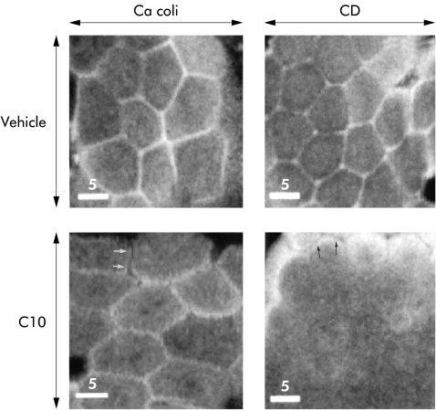
Figure 3.
Perijunctional filamentous (F)-actin distribution in human ileal mucosa. Confocal en face sectioning at the apical level of enterocytes in the villus region in specimens stained with rhodamine-phalloidin. Graphs show specimens from a colon cancer patient (left) and non-inflamed mucosa from a Crohn's disease patient (right) after vehicle experiments (Vehicle; top) and after exposure to sodium caprate (C10; bottom). In the vehicle experiments, both groups showed F-actin arrayed in perijunctional rings. C10 exposed specimens demonstrated reorganisation of F-actin with marked differences in specimens from colon cancer patients and Crohn's disease patients, respectively. In cancer coli specimens (lower left), a more fragmented appearance of the perijunctional rings was seen, with occasional separation of the actin in adjacent cells (arrows). In non-inflamed ileum from Crohn's disease, F-actin staining was diminished at the junctional level (lower right), with staining seen only in patchy small areas. At the level of the microvilli, the F-actin from adjacent cells was separated after C10 exposure (arrows). Bars indicate 5 μm.

