Figure 3.
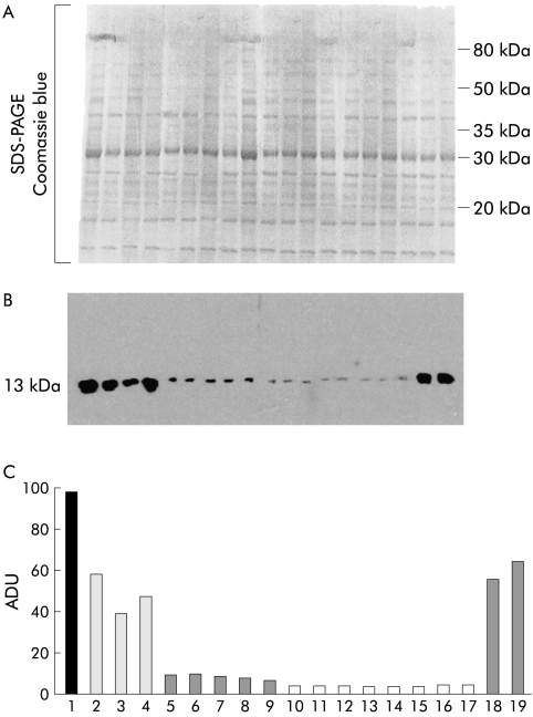
Western blot analysis of transforming growth factor β1 (TGF-β1) protein in patients affected by colon adenocarcinoma at different stages of malignancy. A representative immunoblot. Double protein aliquots (40 μl each) were taken from biopsy specimens after homogenisation. One aliquot was separated by sodium dodecyl sulphate-polyacrylamide gel electrophoresis (SDS-PAGE) and stained with Comassie blue to verify actual protein normalisation (A). The second protein aliquot was also separated by SDS-PAGE but this time processed with a TGF-β1 polyclonal antibody (B). Densitometric analysis of the cytokine level of the single specimens is reported in terms of arbitrary densitometric units (ADU) (C). Samples: 1, colon mucosa from Crohn's disease patient; 2–4, normal colon tissue; 5–9, mucosa from colon adenocarcinoma at T2 (G2) stage; 10–17, mucosa from colon adenocarcinoma at stage T3 (G2); 18–19, mucosa from colon adenocarcinoma at stage T4 (G3).

