Figure 1.
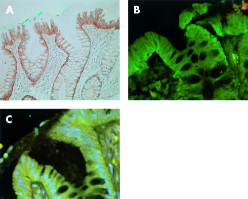
Cadherin and catenin expression in aberrant crypt foci. (A) P-cadherin was expressed in cytoplasm and along cell boundaries, as demonstrated by immunohistochemistry (aberrant crypt marked with green dye) (magnification ×100). (B) Immunofluorescence image using FITC labelled E-cadherin and Texas Red labelled P-cadherin demonstrating general preserved membranous E-cadherin reactivity with cytoplasmic P-cadherin reactivity (magnification ×200). (C) Immunofluorescence image using FITC labelled β-catenin, Texas Red labelled P-cadherin, and 4,6-diaminido-2-phenylindole (DAPI) stained nuclei. Image shows loss of β-catenin from cell membranes with corresponding cytoplasmic/perinuclear staining (pale blue nuclei) and extensive cytoplasmic P-cadherin staining (yellow) (magnification ×200).

