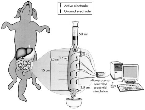
Figure 1.
Schematic diagram of the implantation of four sets of longitudinal subserosal electrodes (15×0.25 mm) at 3 cm intervals. The electrode sets were attached to a multichannel stimulator controlled by specially designed software on a personal computer. A 15 cm segment of descending colon was filled with 40–50 ml of viscous material plus 50 pellets through a transversal opening made 2.0 cm proximal to the first set of electrodes and maintained closed with a purse string suture. The colon was transected 2.5 cm distal to the last set of electrodes. The resulting distal stoma was positioned in a metallic dish to collect eventual emptied material during stimulation and non-stimulation sessions.

