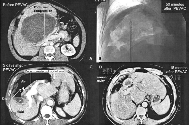
Figure 3.
Computed tomography (CT) scan before percutaneous evacuation (PEVAC) showing portal vein bifurcation compression by “mother and daughter” cysts (A). (B) Cyst cavity 50 minutes after partial evacuation of cyst content. (C) CT scan two days after PEVAC showing decompression of portal vein bifurcation and air fluid level in a non-collapsed cavity. (D) CT scan 18 months after PEVAC showing the remnant cavity.

