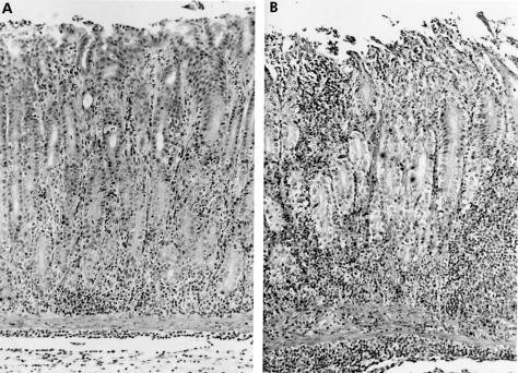
Figure 2.
Microscopic features of the pyloric mucosa in gerbils. (A) Helicobacter pylori group. Moderate amounts of neutrophils and mononuclear cells were seen three weeks after inoculation of H pylori. (B) H pylori and aspirin group. Numerous neutrophils and mononuclear cells as well as deep erosions were evident (haematoxylin and eosin stain, ×100).

