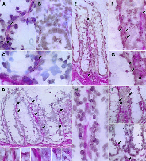
Figure 1.
Y chromosome detection in the mouse. The Y chromosome is seen as a punctate brown/black density in the cell nucleus. Red staining of the cell cytoplasm identifies positive immunohistochemical staining. (A) Male positive control showing Y chromosome detection in colonic pericryptal myofibroblast cells immunostained with the α smooth muscle actin (αSMA) antibody. (B) Female control demonstrating lack of Y chromosome detection within colonic pericryptal myofibroblasts immunostained with the αSMA antibody. (C–G) Female mouse after male whole bone marrow transplantation showing Y chromosome positive myofibroblasts immunostained with the αSMA antibody. (C) Few αSMA immunoreactive Y chromosome positive cells present in the mouse colon seven days after male bone marrow transplant. (D) Numerous Y chromosome positive myofibroblasts (for example, arrows, see also inserts for high power views) immunoreactive for αSMA in female mouse colon 14 days after male bone marrow transplant. (E) Y chromosome positive myofibroblasts stained with αSMA in the colon at six weeks after transplant showing extension of the column right up to the top of the crypts. (F, G) High power view of Y chromosome positive myofibroblasts that are αSMA immunoreactive in female mouse colon six weeks after transplant. (H) Desmin positive cells within the lamina propria were not Y chromosome positive. (I) Y chromosome positive cells that are negative for the haematopoeitic progenitor marker CD34 in the mouse colon six weeks after transplant. (J) Y chromosome positive cells that are negative for the mouse macrophage marker F4/80 antigen (positively stained macrophages shown by arrows).

