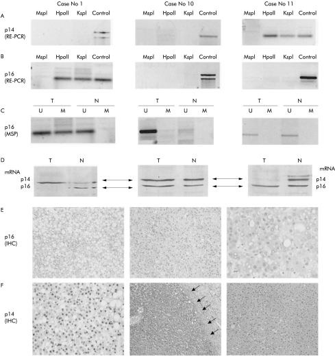
Figure 1.
Analysis of p14ARF and p16INK4a in three liver cell adenomas (case Nos 1, 10, and 11; same patients as in table 1 ▶). (A) p14ARF analysis with restriction enzyme related-polymerase chain reaction (RE-PCR). The methyl sensitive restriction enzymes used for RE-PCR are indicated (HpaII, KspI); digestion with the non-methyl sensitive enzyme MspI serves as a negative control and undigested DNA (control) serves as a positive control. The p14ARF gene is methylated in case No 11 and unmethylated in case Nos 1 and 10. (B) p16INK4a analysis with RE-PCR. Similar to (A), the methyl sensitive restriction enzymes used for RE-PCR are indicated (HpaII, KspI); digestion with the non-methyl sensitive enzyme MspI serves as a negative control and undigested DNA (control) serves as a positive control. Methylation of p16 INK4a is detected in case No 1, but not in case Nos 10 and 11. (C) p16 INK4a analysis using methylation specific polymerase chain reaction (MSP). Bisulphite treated DNA (which changes the unmethylated but not the methylated cytosines into uracil) is subjected to PCR amplification using primers designed to anneal specifically to the methylated bisulphite modified DNA. MSP results are expressed as unmethylated p16 specific bands (U) or methylated p16 specific bands (M). Bisulphite converted DNA from normal corresponding liver tissue (N) served as a negative control, as indicated by the presence of the U but not the M band. Similar to (B), methylation of p16 INK4a was detected in case No 1 but not in case Nos 10 and 11. (D) Results of multiplex reverse transcription-PCR (RT-PCR) of p14 mRNA (upper line corresponding to 200 bp) and p16 mRNA (lower line corresponding to 179 bp) for case Nos 1, 10, and 11. (E) Immunostaining of p16 INK4A protein in liver cell adenoma (LCA). Case No 1 shows methylated p16 INK4a and complete loss of p16 INK4a (LCA cells negative for p16 protein) (original magnification ×10). p16 INK4a is detectable in case Nos 10 and case 11 (dark reaction product within the cell nuclei) (original magnification ×20 and ×40). (F) Immunostaining of p14ARF protein in LCA. Case No 1 shows unmethylated p14 ARF and strong immunoreactivity of the tumour cells for p14 protein (dark reaction product within the tumour cell nuclei) (original magnification ×40). Case No 10 with unmethylated p14 ARF and strong immunoreactivity of the tumour cells for p14 protein (dark reaction product within the tumour cell nuclei). The tumour surrounding fibrous capsule (arrows) is negative (original magnification ×5). Case No 11 shows a methylated p14ARF and complete protein loss within the tumour tissue (original magnification ×20).

