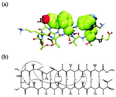
Figure 1.
Native hairpin structure. (a) Space-filling model of the hydrophobic cluster for the minimized coordinates extracted from the NMR structure of the complete Ig-binding domain of protein G (Protein Data Bank code 2gb1) (13). (b) Schematic. Backbone hydrogen bonds contributing to the sheet are indicated by dashed lines and numbered. In general, we count a hydrogen bond if the corresponding heavy atoms are within 3.4 Å of each other and the out-of-line angle (180°− ∡DHA) is less than 70°. Arrows indicate the pairwise distances used in the cluster analysis: D47H–A48H, A48H–T49H, T49H–K50H, K50H–T51H, W43Cα–V54Cα, Y45Cα–F52Cα, W43Cɛ3–F52Cβ, Y45Cβ–F52Cγ, and D47Cα–F52Cγ.

