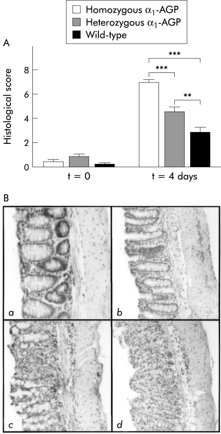
Figure 3.
(A) Histological score after dextran sodium sulphate (DSS) treatment. Four days after 2% DSS treatment, the histological score was determined from the distal part of the colon from homozygous α1-AGP-transgenic (n=18), heterozygous α1-AGP-transgenic (n=19), and wild-type mice (n=15) and compared with negative controls of the corresponding genotype (n=5 for each group). Statistical significance values are based on homozygous and heterozygous transgenic versus wild-type mice, and homozygous versus heterozygous transgenic mice: **p<0.01, ***p<0.001. (B) Representative distal colon sections (100×) stained with haematoxylin-eosin. (a) Section of a negative control (no DSS treatment) showing normal crypt morphology, with crypt bases resting on the lamina muscularis mucosa, and no inflammatory infiltrate. (b) Section of a wild-type mouse four days after DSS treatment. There is local loss of goblet cells and some inflammatory infiltrate at the crypt base which is no longer resting on the lamina muscularis mucosa. (c) Section of a heterozygous transgenic mouse showing local crypt destruction and inflammatory infiltrate reaching to the lamina muscularis mucosa. (d) Section of a homozygous transgenic mouse with total destruction of crypt structure and inflammatory infiltrate reaching into the submucosa.

