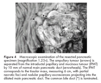
Figure 4.

Macroscopic examination of the resected pancreatic specimen (magnification 1.25×). The ampullary tumour (arrows) is separated from the intraductal papillary and mucinous tumour (IPMT) by 10 mm of normal main pancreatic duct (arrowheads). The IPMT corresponds to the tissular mass, measuring 6 cm, with partial necrotic foci and nodular papillary excrescences projecting into the dilated main pancreatic duct. The common bile duct (*) is laminated.
