
Figure 5.
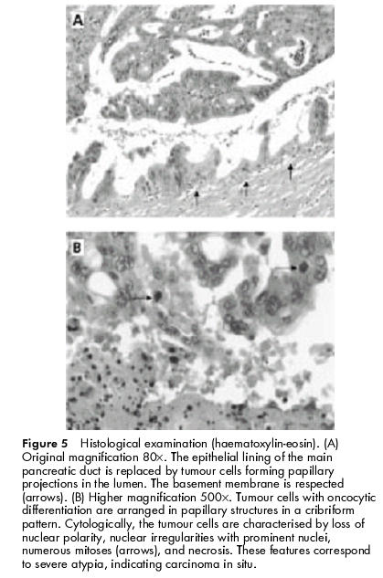
Histological examination (haematoxylin-eosin). (A) Original magnification 80×. The epithelial lining of the main pancreatic duct is replaced by tumour cells forming papillary projections in the lumen. The basement membrane is respected (arrows). (B) Higher magnification 500×. Tumour cells with oncocytic differentiation are arranged in papillary structures in a cribriform pattern. Cytologically, the tumour cells are characterised by loss of nuclear polarity, nuclear irregularities with prominent nuclei, numerous mitoses (arrows), and necrosis. These features correspond to severe atypia, indicating carcinoma in situ.