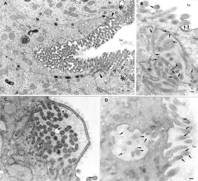
Figure 4.
Epon sections of small bowel biopsies from patients with microvillus inclusion disease demonstrating autophagocytosis of the apical membrane of the villus enterocyte. The biopsies shown in (B) and (C) were incubated with cationised ferritin (arrows) for five minutes. (A) Engulfing of the apical membrane deeply inwards in the enterocyte. Arrowheads indicate the zone between the apical and basolateral membranes and the asterisks the apical region of the neighbouring enterocytes. (B) Cationised ferritin appears to be attached to the membranes of microvilli contained within a microvillus inclusion body which was located in the vicinity of the apical membrane. (C) Infolded microvilli covered by a thin cytosolic strand. The structure contains two vesicles with cationised ferritin particles on their membranes (arrows). (D) Cationised ferritin (arrows) is not only detectable on microvilli but also within vesicles of an invagination of the apical membrane. D, desmosome; Lu, intestinal lumen; Mi, microvilli; bars=0.1 μm.

