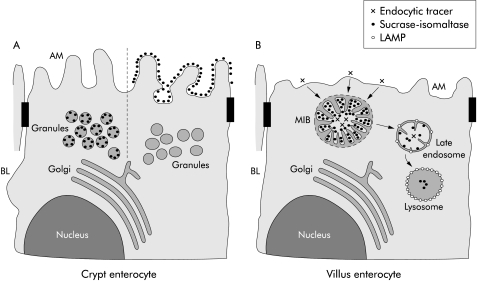
Figure 6.
A model of the exocytic and endocytic pathways in crypt and villus microvillus inclusion disease (MID) epithelial cells. (A) Secretory granules, which are present in crypt epithelial cells, are labelled by the sucrase-isomaltase (SI) antibody with variable intensity. Crypt epithelial cells whose secretory granules do not contain SI show SI labelling on the apical membrane (AM). In contrast, crypt epithelial cells with no SI labelling on microvilli reveal secretory granules with SI staining. (B) Villus enterocytes with detectable microvillus inclusion bodies (MIBs) are characterised by a few and shortened microvilli. Microvilli within MIBs are labelled by the SI antibody but are negative for lysosome associated membrane protein (LAMP) indicating their early endosomal nature. Late endosomes of MID enterocytes, which are identified by LAMP positivity, reveal few microvilli-like projections and SI labelling. Lysosomes, which are strongly positive for LAMP, contain a low amount of SI without any microvilli-like structures. BL, basolateral membrane.

